Nail anatomy: essential parts of the nail every nail technician should know
By Katie Barnes | 27 March 2025 | Expert Advice, Feature

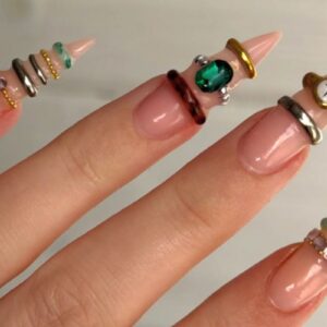
As a nail professional, it is paramount that you familiarise yourself with all parts of nail anatomy to ensure you are offering your clients a professional and thorough service.
Understanding the purpose and growth of the nail
The primary function of the nail is to protect the outermost bone of the finger or toe, the fingertip and the surrounding soft tissues from injuries.
The fingertips contain a lot of sensory endings that allow us to distinguish touch, pain and temperature. Since these nerve fibres are highly sensitive and fragile, it is crucial to protect them.
The underside of the finger is fleshy, while the nail covers the pad-like topside. The nail plate is designed to crack or break off under pressure or impact, acting as a shock absorber so the nail bed and nerve endings experience minimal damage.
The cells of the nail originate in the matrix and move forward towards the fingertips. When they first emerge from under the cuticle, they are soft and spongy. They only harden and keratinise when exposed to the air: a process which usually takes several days.
The nail continues to grow forward in the shape and width of the nail bed. When the nail plate extends beyond the hyponychium, this becomes the free edge. It is important to have a free edge, as this ensures that all the nail bed is covered and protected.
Let’s explore the different parts of the nail:
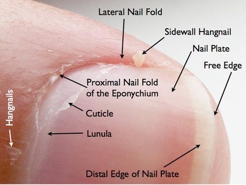
Cuticle
The cuticle is the strip of hardened, flaky skin found on the nail plate, above the eponychium, at the base of a nail.
Eponychium
What most people consider the cuticle is actually the living tissue known as the eponychium. It serves as a barrier to prevent pathogens from entering the soft tissue. The skin fold ends at the base of the nail plate, and is important to be treated with care, as infection can occur if the matrix seal is broken.
Hyponychium
The hyponychium is the tissue under the free edge of the nail that seals the nail plate to the tip of the finger. It acts as a barrier to prevent pathogenic bacteria from entering the finger. Some clients may have a condition called ‘extended hyponychium’, where this skin protrudes beyond the nail plate and tip of the finger. You must treat these clients’ nails with care, as this can become painful if the nail is filed too short.
Nail bed
The nail bed is located under the nail plate. It starts at the base of the nail (matrix) and extends to the free edge. Like all skin, it is made up of dermis and epidermis. The nail bed contains thousands of blood vessels that carry food, oxygen and nutrients to the fingernail.
Nail plate
The nail plate is what we commonly refer to as ‘the nail’. It has a densely packed surface made of keratin. Several layers of dead, compacted cells give the nail its strength and flexibility. These cells are not living; they are keratinised.
The pink appearance of the nail comes from the blood vessels beneath it, as the nail plate itself is translucent. The underside of the nail plate has grooves along the length that help anchor it to the nail bed and ensure the nail grows in the correct direction. The purpose of the nail plate is to protect the living nail bed underneath.
Lanula
The lanula is the visible part of the matrix that resembles a half moon. It should be treated with care as the cells in this area have not yet fully keratinised. The lanula is white in colour and opaque.
Nail folds (lateral & proximal)
The nail folds protect the nail matrix. The proximal and lateral nail folds are part of the skin, which doesn’t end at the nail edge but continues beneath it in folds. The continuing skin acts as a protective barrier, sealing the matrix from bacteria and dirt that is commonly found within our environment.
Nail matrix
The matrix is the root of the nail. This area is not visible, as it is hidden and protected by the proximal nail fold. The matrix produces keratin cells that make up the nail plate. As new cells are produced, older ones are pushed outwards and flattened. This process causes the cells to lose their original white, plump appearance and eventually become transparent, becoming part of the nail plate.
The width and thickness of the nail plate is determined by the size, length and thickness of the matrix, while the shape of the fingertip determines if the nail plate is flat, arched or hooked. The matrix is the most important feature of the nail unit. Damage to the matrix can cause permanent damage to the appearance of the nail.
Love Katie x
*Originally published November 2017

Read the latest issue